Ciliary Ganglion - Autonomic control of the eye | Anatomy Tutorial
71.3 هزار بار بازدید -
4 سال پیش
-
#ciliary
#ciliary #eye #oculomotor
Link for Donations https://paypal.me/studentlamedicina?l...
Instagram: anatomy.knowledge
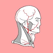
The ciliary ganglion in humans is approximately 3mm in size, and located approximately 5 mm posterior to the globe and lateral to the optic nerve. It is a parasympathetic ganglion and contains approximately 2500 neurons.
From the forepart of the ganglion are arising the short cilliary nerves.They pierce the sclera at the back part of the bulb of the eye.
The cilliary ganglion has three roots.
The sensory root arises from the nasociliary nerve, a branch of the ophthalmic nerve and unites with the cilliary ganglion at its posterior superior angle. The sensory root transmits preripheral projections from the pseudouniporal neurons located in the trigeminal ganglion. These fibers pass through the cilliary ganglion and further on piercing the eye by way of short cilliary nerves , they travel in the pericoroidal space to supply the cornea, cilliary body and iris. Some of these peripheral projections from the trigeminal ganglion follow the course of the long cilliary nerves , branches of the nasociliary nerve, which pierce the posterior part of the eyeball.
The second root of the cliary ganglion to be described is the motor root. The motor root is a branch of the nerve to inferior oblique muscle and unites with the ciliary ganglion at it posterior inferior angle. The nerve to inferior oblique muscle is a branch of the inferior division of the oculomotor nerve. Parasympathetic preganglionic fibers from edinger-westphal nucleus located in the midbrain will travel within the oculomotor nerve, then within its inferior division and arrive at the ciliary ganglion by way of its motor root. In the ciliary ganglion the parasympathetic preganglionic fibers will sinapse with the parasympathetic neurons located within the ganglion. From the cilliary ganglion the parasympathetic postganglionic fibers will pierce the posterior end of the eyeball by way of short ciliary nerves. Traveling in the pericoroidal space, these parasympathetic fibers will supply the cilliary muscle and pupilar sphincter muscles. Stimulation of these fibers constricts the pupil and accommodates the eye for near vision.
The third root of the cilliary ganglion is the sympathetic root which contains sympathetic postganglion fibers.
Link for Donations https://paypal.me/studentlamedicina?l...
Instagram: anatomy.knowledge
The ciliary ganglion in humans is approximately 3mm in size, and located approximately 5 mm posterior to the globe and lateral to the optic nerve. It is a parasympathetic ganglion and contains approximately 2500 neurons.
From the forepart of the ganglion are arising the short cilliary nerves.They pierce the sclera at the back part of the bulb of the eye.
The cilliary ganglion has three roots.
The sensory root arises from the nasociliary nerve, a branch of the ophthalmic nerve and unites with the cilliary ganglion at its posterior superior angle. The sensory root transmits preripheral projections from the pseudouniporal neurons located in the trigeminal ganglion. These fibers pass through the cilliary ganglion and further on piercing the eye by way of short cilliary nerves , they travel in the pericoroidal space to supply the cornea, cilliary body and iris. Some of these peripheral projections from the trigeminal ganglion follow the course of the long cilliary nerves , branches of the nasociliary nerve, which pierce the posterior part of the eyeball.
The second root of the cliary ganglion to be described is the motor root. The motor root is a branch of the nerve to inferior oblique muscle and unites with the ciliary ganglion at it posterior inferior angle. The nerve to inferior oblique muscle is a branch of the inferior division of the oculomotor nerve. Parasympathetic preganglionic fibers from edinger-westphal nucleus located in the midbrain will travel within the oculomotor nerve, then within its inferior division and arrive at the ciliary ganglion by way of its motor root. In the ciliary ganglion the parasympathetic preganglionic fibers will sinapse with the parasympathetic neurons located within the ganglion. From the cilliary ganglion the parasympathetic postganglionic fibers will pierce the posterior end of the eyeball by way of short ciliary nerves. Traveling in the pericoroidal space, these parasympathetic fibers will supply the cilliary muscle and pupilar sphincter muscles. Stimulation of these fibers constricts the pupil and accommodates the eye for near vision.
The third root of the cilliary ganglion is the sympathetic root which contains sympathetic postganglion fibers.
4 سال پیش
در تاریخ 1399/06/01 منتشر شده
است.
71,398
بـار بازدید شده