Case of the Week: Perihilar Cholangiocarcinoma/Klatskin Tumor (CT & MRI)
11.7 هزار بار بازدید -
3 سال پیش
-
In this radiology lecture, we
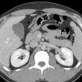
In this radiology lecture, we discuss the CT and MRI appearance of perihilar cholangiocarcinoma.
Key points include:
1) Perihilar cholangiocarcinoma (AKA Klatskin tumor) occurs at bifurcation of the hepatic duct.
2) Cholangiocarcinoma (CC) is a primary malignant tumor of bile duct epithelium, usually adenocarcinoma.
3) CC is the most common primary hepatic malignancy after hepatocellular carcinoma (HCC), and most are extrahepatic (as opposed to intrahepatic).
4) Appearance of CC is based on growth pattern: Mass-forming, periductal infiltrating, and intraductal growing.
5) Risk factors: Parasite infection, choledochal cyst, primary sclerosing cholangitis, recurrent pyogenic cholangitis, and inflammatory bowel disease (ulcerative colitis).
6) Patients are usually 65 or older.
7) On CT and MRI, perihilar CC appears as a biliary stricture with shouldering/abrupt tapering.
8) If a mass is visible, will typically have rimlike enhancement with gradual centripetal enhancement on delayed images, be T2 bright (but not as homogeneous or as bright as hemangioma), and may have a targetlike appearance on DWI (favors CC over HCC)
Click the Community tab or follow on social media for bonus teaching material posted throughout the week!
Website: www.radiologistHQ.com/
Video Podcast: bit.ly/radiologistHQ
Instagram: www.instagram.com/radiologistHQ/
Facebook: www.facebook.com/radiologistHeadQuarters/
Twitter: twitter.com/radiologistHQ
3 سال پیش
در تاریخ 1400/10/15 منتشر شده
است.
11,728
بـار بازدید شده